Personalized Plastic Surgery & Aesthetic Care in Seocho-gu, Seoul

Medical Guide
Mole Removal in Korea: CO2 Laser, Erbium, or Excision
Lesion type and depth, not patient preference, decide whether a mole, milia, wart or scar is treated with CO2 laser, erbium laser or surgical excision.
Schedule a ConsultationScar and Skin Lesion Removal: How the Diagnosis Decides Between CO2 Laser, Erbium Laser, and Excision
Medically reviewed by Dr. In-Ho Khang, MD, Yonsei University College of Medicine; Chief Academic Director of Plastic Surgery, Korean Society of Aesthetic Surgery & Laser (KSASL)
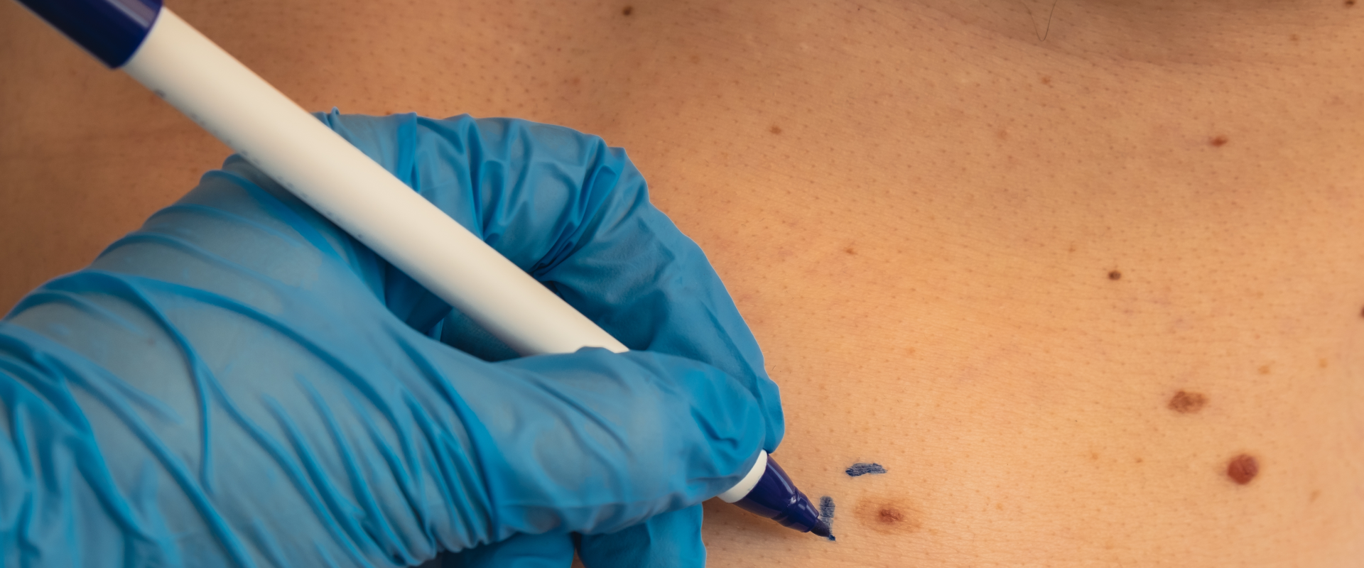
If you are researching what mole removal Korea clinics offer as a short cosmetic add-on to a trip, the thing to understand first is that CO2 laser, erbium laser, fractional resurfacing, and surgical excision are not a menu you choose from. They are conclusions someone reaches after examining the lesion. Its type, its depth, and whether a pathologist needs to see the tissue narrow the options before convenience enters the conversation. YONSEI LEEDS CLINIC in Seoul states that it assesses each lesion clinically before deciding whether a laser is appropriate at all.
Why the Order of Operations Decides Everything Else
A removal request usually arrives as a cosmetic question: this bump bothers me, can it go. The clinical answer starts elsewhere, because the same visible bump can be a benign mole, a seborrheic keratosis, a skin tag, a small cyst, a viral infection, or something that needs a laboratory to identify. Those are not variations of one problem. They sit at different depths and respond to different mechanisms: removing a keratin cyst is a mechanical job, clearing flat warts is an antiviral job, flattening a raised nevus is an ablative job, and improving a depressed scar is a remodeling job that takes months.
So the sequence runs diagnosis, then depth, then modality, then settings. Reversing it is how people end up with an incompletely removed lesion, a mark where the lesion used to be, or a recurrence six months later. The clinic states that no laser is applied before that assessment is complete.
What Is Actually Sitting on Your Skin
Patients rarely arrive with a diagnosis, and a great many "moles" are not moles.
Melanocytic Nevi
A mole in the ordinary sense is a cluster of pigment-producing cells. The National Library of Medicine describes moles as growths that occur when melanocytes cluster and notes that most people have between 10 and 40. The National Cancer Institute adds that common moles are usually smaller than 5 millimeters, that dysplastic nevi are usually larger, and that having more than five dysplastic nevi is associated with roughly ten times the melanoma risk of having none. Nevi also vary in depth: some sit largely in the epidermis, others extend well into the dermis — a difference invisible from the surface that determines whether an ablative laser can reach the whole thing.
Seborrheic Keratoses, Skin Tags, and Sebaceous Hyperplasia
These are the three things most often mistaken for moles. Seborrheic keratoses are benign epidermal growths with a waxy, "stuck-on" look; clinical references describe them as near-universal in adults over 60 and note that both erbium and CO2 lasers have been used to remove them. Skin tags are small pedunculated growths in areas of friction. Sebaceous hyperplasia produces soft yellowish papules with a central dimple. All three are superficial or near-superficial. YONSEI LEEDS CLINIC lists all of them, along with milia, flat warts, syringomas, post-acne scars, and keloids, among the concerns it assesses.
Milia Are Cysts, Not Spots
Milia are often treated as though they were whiteheads, which is why they often do not resolve. They are small keratin cysts sitting beneath the epidermis, typically under 3 millimeters, and present in roughly 40 to 50 percent of healthy full-term newborns before clearing on their own. In adults they persist.
Secondary milia often arise from sweat ducts and are documented as following dermabrasion, burns, skin grafting, and radiotherapy. Because the contents are keratin inside a closed sac, standard management is to open the sac and express the contents, with topical retinoids or electrodesiccation as alternatives. A resurfacing pass over the surface does not reliably solve a cyst sitting beneath it — and milia are themselves a recognized complication of ablative resurfacing.
Flat Warts Are an Infection
Verruca plana, or flat warts, are caused by human papillomavirus, most often types 3, 10, and 28. They typically measure 1 to 7 millimeters and can appear in dozens or hundreds, often spreading along lines where skin has been shaved or scratched. Roughly two-thirds of warts in general resolve on their own within 12 to 24 months.
Destroying one wart does not treat virus in the surrounding skin, which is why flat warts characteristically need several sessions and can recur. The clinic states that flat warts and scar conditions generally require multiple sessions, while discrete lesions such as moles, milia, and skin tags may be resolved in one session when correctly diagnosed and treated at the right depth.
The Evaluation That Comes Before Any Cosmetic Removal
A pigmented lesion you want removed for appearance is still a pigmented lesion, and it should be evaluated on its own merits first.
The American Academy of Dermatology's ABCDE criteria are the standard shorthand: asymmetry, an irregular or poorly defined border, color varying from one area of the spot to another, diameter — melanomas are usually larger than about 6 millimeters at diagnosis, though they can be smaller — and evolution, meaning the spot is changing or simply looks unlike your others. The National Library of Medicine puts it more briefly: have a lesion checked if it looks unusual, grows, or changes in color or outline.
Examination by eye is not the whole picture. A meta-analysis of nine studies covering 8,487 suspicious lesions and 375 melanomas found that clinical examination combined with dermoscopy reached a sensitivity of about 0.90, against about 0.71 for naked-eye examination alone. Asking whether your lesion will be looked at with a dermatoscope is a reasonable, specific question.
One point from the National Cancer Institute cuts against a common assumption: removing a normal mole as a preventive measure is not recommended. Taking a mole off does not lower melanoma risk.
Why Destroying a Lesion Also Destroys the Evidence
Diagnosis of melanoma requires tissue. The National Cancer Institute states that the only way to diagnose it is to remove tissue and have a pathologist examine it. A laser that ablates a lesion does not produce that tissue in usable form. The lesion is gone, and so is the answer.
Guidance on technique is correspondingly firm. The National Cancer Institute's professional-level melanoma summary states that a biopsy, preferably by local excision, should be performed for any suspicious lesion, and adds that "Suspicious lesions should never be shaved off or cauterized." The National Library of Medicine draws the same line: an excisional biopsy removes the entire lesion including deep skin and fat and is most commonly performed when melanoma is suspected, while a shave biopsy takes only the outermost layers.
The reason is depth. Breslow thickness — how far a melanoma extends into the skin — drives staging and therefore treatment, and clinical references note that transection at the base of a specimen can compromise the accuracy of that measurement. A systematic review and meta-analysis of biopsy technique in cutaneous melanoma found that guidelines across Australia, Europe, the United Kingdom, and the United States converge on full-thickness excision with narrow lateral margins of roughly 1 to 3 millimeters as the diagnostic procedure of choice.
There is also a failure mode specific to cosmetic light-based treatment. A case report in a peer-reviewed family medicine journal describes a facial "sunspot" treated with intense pulsed light in a non-medical setting; the area later repigmented and grew, and biopsy showed lentigo maligna melanoma requiring wide local excision and flap reconstruction. The authors make the general point directly: unlike excisional biopsy, light-based treatment does not permit histologic evaluation, so it gives neither a tissue diagnosis nor margin assessment.
None of this means a mole you dislike must be surgically excised. It means the decision belongs to someone who has examined it.
CO2 at 10,600 Nanometers Versus Erbium at 2,940 Nanometers
Both are ablative lasers: both remove tissue by vaporizing the water inside it. The difference between them is almost entirely how strongly water absorbs their wavelength. The CO2 laser emits at 10,600 nanometers. The erbium-doped yttrium aluminum garnet laser, erbium:YAG, emits at 2,940 nanometers, close to the water absorption peak around 3,000 nanometers. Clinical references put erbium's absorption by water at roughly 12 to 18 times that of CO2. Three consequences follow:
- Ablation per pass. A first pass with a CO2 laser vaporizes on the order of 20 to 60 micrometers of tissue; a pass with erbium removes roughly 3 to 5 micrometers. CO2 works in larger increments, erbium in thin controllable layers.
- Residual thermal damage. Because erbium's energy is absorbed almost entirely at the surface, less heat spreads sideways. Clinical references describe a residual thermal damage zone around 10 to 40 micrometers for erbium against roughly 100 to 150 micrometers for CO2. A micrometer is a thousandth of a millimeter — small distances, but they set how much inflammation the surrounding skin experiences.
- Bleeding and collagen. That thermal spread is not purely a liability. Heat from CO2 seals small vessels and contracts collagen, which is useful in some raised lesions. Erbium's cleaner ablation gives less of both, and clinical references report faster re-epithelialization after erbium resurfacing than after CO2.
That is a decision rule, not a ranking. CO2 tends to suit raised and textured lesions where tissue must be removed in depth and some coagulation helps; erbium tends to suit shallower lesions and situations where limiting heat delivered to surrounding skin is the priority. YONSEI LEEDS CLINIC describes the same split.
What Thermal Spread Means for Skin That Pigments Easily
Those thermal damage figures are not trivia. Heat deposited around a treatment site produces inflammation, and inflammation is a documented trigger for post-inflammatory hyperpigmentation — darkening that follows the injury rather than the original lesion.
Clinical references describe it as more common and more severe in Fitzpatrick skin types III through VI, list laser and light therapy explicitly among its causes, and note that pigment confined to the epidermis typically resolves over 6 to 12 months while pigment deposited in the dermis may be effectively permanent. In one scar-resurfacing report covering Fitzpatrick III to V patients, 45.5 percent developed transient hyperpigmentation, typically two to four weeks after treatment, resolving within three months in about 90 percent of those affected.
This is why parameters are skin-specific, not lesion-specific alone. Clinical references advise against non-fractional CO2 resurfacing in Fitzpatrick IV and above, and recommend that types III and IV be treated fractionally and only after a test spot has healed for two to three weeks. Those cautions concern broad resurfacing rather than precise ablation of one small lesion, but the biology is the same. The clinic states that laser depth and settings are calibrated to individual skin type specifically to address this risk, and notes that ultraviolet exposure during healing can increase the chance of pigmentation change.
Fractional Resurfacing Solves a Different Problem
Everything above concerns removing something discrete. Scars are the opposite problem: the tissue is already there, and the goal is to reorganize it.
Fractional ablative resurfacing creates narrow microscopic columns of thermal injury separated by untreated skin. Clinical references describe these columns as under 400 micrometers across and reaching as deep as 1,300 micrometers, with surrounding intact tissue serving as a reservoir for healing. That allows deeper treatment than full-field ablation with shorter recovery. Results are real but partial: one reference reports fractional CO2 producing meaningful improvement in about 70 percent of atrophic traumatic scars over roughly six monthly sessions. The American Academy of Dermatology is blunter — laser treatment cannot eliminate a scar; it replaces a noticeable scar with a less noticeable one, and it advises against treating tanned or sunburned skin.
Raised scars behave differently again. The American Academy of Dermatology describes hypertrophic scars as appearing one to two months after injury, staying within the boundaries of the original wound, and often fading — while keloids grow beyond the original wound over months or years and do not fade on their own. It notes that between roughly a third and half of people who develop keloids have at least one blood relative who does, and clinical references on scar revision report keloid incidence in the range of 4 to 16 percent in African-American, Hispanic, and Asian populations. If keloids run in your family, say so before any lesion is removed anywhere. The clinic states that fractional CO2 is used for acne scars, hypertrophic scars, and textural irregularities.
Excision, and the Scar You Accept in Exchange
Surgical excision trades a lesion for a line. It also offers two things no laser can: a specimen for the pathologist, and removal of a lesion's full depth in one step.
The National Library of Medicine describes the experience plainly: a punch biopsy takes an area about the size of a pencil eraser, stitches typically come out between three and fourteen days later, and the recognized risks are infection and scarring or keloid formation.
Excision is the right answer when a lesion is deep, atypical, requires histologic assessment, or when a previous destructive treatment left something behind. The clinic states that surgical excision with biopsy is available for those situations, and that patients with complex lesions or previous incomplete treatment can access scar revision capability. If a lesion needs a diagnosis, the line is not an optional cost — it is the procedure.
What YONSEI LEEDS CLINIC States About Its Own Sequence
The clinic publishes a five-step process for scar and spot removal: clinical evaluation of each lesion for type, depth, border characteristics, and suitability for laser; identification of atypical lesions, with cases requiring further assessment referred for biopsy; treatment selection among CO2, erbium, fractional CO2, and surgical excision; treatment planning covering expected outcomes, recovery, risks, and anticipated sessions; and follow-up to evaluate healing.
On the procedure itself, the clinic states that topical anesthetic cream may be applied for roughly 20 to 40 minutes where appropriate, that treatment is followed by a healing ointment or protective dressing, and that patients receive instructions on wound hygiene, activity restriction, and sun avoidance. It describes most individual lesion removals as taking minutes, with larger areas or scar-resurfacing sessions running approximately 20 to 45 minutes. Its stated position on outcomes is careful: results depend on lesion type, healing response, and skin type, and scarring or pigmentation changes cannot be completely ruled out.
The clinic describes its approach as combining plastic surgery and dermatology expertise, and Dr. In-Ho Khang is identified as Representative Director of Yonsei Leeds Plastic Surgery & Dermatology Clinic. That is the clinic's own description of how it organizes its practice, not a comparative claim. Dr. Khang holds an MD from Yonsei University College of Medicine and serves as Chief Academic Director of Plastic Surgery at the Korean Society of Aesthetic Surgery & Laser; his doctor profile page is the clinic's published record of that background. The clinic describes him as a board-certified specialist without naming the issuing board, so treat that as the clinic's statement and ask to see the certificate.
What the Clinic Does Not Publish
Reading a clinic's documentation honestly includes noticing the blanks. For this treatment, several fields are marked as needing confirmation rather than answered: who is not a suitable candidate, the risks a patient should know, the recovery timeline, how long a session takes, which anesthetic is used, what to avoid beforehand, and anything about cost. Case volumes, years in practice, booking lead times, payment options, and travel logistics are unpublished as well.
A blank field is more careful than an invented one, but those answers have to come from your consultation. Ask specifically: what is this lesion, how will you confirm it, what happens to the tissue, and what will the site look like at one week and at three months.
Planning Removal Around a Trip to Seoul
Lesion removal is short, which makes it tempting to slot in at the end of a trip. Two things argue for the opposite. First, follow-up: the clinic states that follow-up visits assess healing, confirm removal, and decide whether further treatment is needed, and if a lesion warrants excision and histology, both a result and a wound check take time. Second, flying: the US Centers for Disease Control and Prevention's medical tourism guidance cites plastic surgery society advice to wait 7 to 10 days after laser treatments or facial cosmetic procedures before flying, and identifies infection as the most common complication reported in medical travel.
The clinic's published hours are Monday through Friday, 10:30 AM to 8:00 PM, and Saturday, 10:00 AM to 4:00 PM, closed Sundays and public holidays. It is on the fifth floor of Il-i Tower in Seocho-gu, roughly one minute from Gyodae Station Exit 12. Seoul runs roughly 13 to 17 hours ahead of the continental United States, which matters when arranging a call. The clinic states that English-speaking consultations, online consultation before traveling, and remote follow-up after returning home are available; its inquiry and consultation page is the published route for arranging either, and its main website carries location information. Send clear, well-lit photographs in advance, and say plainly whether the lesion has changed.
The Bottom Line
- Lesion type and depth determine the modality: CO2 suits raised or textured lesions, erbium suits shallower work with less thermal spread, fractional resurfacing addresses scars, and excision is for deep, atypical, or histology-requiring lesions.
- Any pigmented lesion should be evaluated before it is removed for appearance, because ablating or shaving it destroys the tissue a pathologist would need; the National Cancer Institute advises excisional biopsy for suspicious lesions and warns against shaving or cauterizing them.
- Water absorbs erbium:YAG at 2,940 nanometers roughly 12 to 18 times as strongly as CO2 at 10,600 nanometers, so erbium ablates in thinner layers and leaves a smaller zone of residual thermal damage — which matters most in skin prone to post-inflammatory hyperpigmentation.
- Milia are keratin cysts and flat warts are a viral infection; both need mechanisms different from ablating a surface.
- YONSEI LEEDS CLINIC publishes a diagnosis-first sequence but not its contraindications, risks, recovery timelines, session lengths, or cost, so ask for those at consultation.
Frequently Asked Questions
Should I have a mole checked before having it removed for cosmetic reasons?
Yes. Diagnosis of melanoma requires tissue examined by a pathologist, and destroying a lesion with a laser eliminates that possibility. The National Cancer Institute recommends biopsy, preferably by local excision, for any suspicious lesion and advises against shaving or cauterizing such lesions. Ask what your lesion is and how that was determined before agreeing to a removal method.
What is the difference between CO2 and erbium lasers for skin tag removal Seoul clinics perform?
Both vaporize tissue, but water absorbs erbium's 2,940-nanometer wavelength roughly 12 to 18 times as strongly as CO2's 10,600 nanometers. Erbium therefore removes thinner layers per pass and leaves less residual heat in surrounding tissue; CO2 removes more per pass and produces more coagulation, which helps with raised lesions. The choice follows the lesion.
Will one session remove my mole, or will I need to come back?
It depends on what the lesion is and how deep it goes. YONSEI LEEDS CLINIC states that discrete lesions such as moles, milia, and skin tags may be resolved in a single session when correctly diagnosed and treated at the appropriate depth, while flat warts and scar conditions generally require several. A lesion extending deep into the dermis is more likely to recur if treated too superficially.
Is the CO2 laser vs surgical excision scar trade-off always better with the laser?
Not always. Laser removal usually avoids a linear scar but can leave a flat mark, altered pigment, or an incompletely removed lesion if depth was underestimated. Excision leaves a planned line but removes full depth and produces a specimen for histology. Where the lesion sits, how deep it is, and whether a diagnosis is needed decide which trade-off makes sense.
Why do my milia keep coming back after facials?
Milia are small keratin cysts sitting beneath the surface rather than blocked pores, which is why surface treatments often fail. Standard management is opening the cyst and expressing its contents, with topical retinoids or electrodesiccation as alternatives. Secondary milia are also documented as arising after skin injury, including after resurfacing procedures.
How likely is darkening after laser removal on darker skin?
Post-inflammatory hyperpigmentation is more common and more persistent in Fitzpatrick types III through VI, and laser treatment is a recognized trigger. In one scar-resurfacing report covering types III to V, transient hyperpigmentation occurred in around 45 percent of patients, resolving within three months in most cases. Epidermal pigment typically fades over 6 to 12 months; dermal pigment may not.
How soon can I fly home after having a lesion removed?
Ask the clinic, because it depends on what was done. Medical travel guidance from the US Centers for Disease Control and Prevention cites plastic surgery society advice to wait 7 to 10 days after laser treatments or facial cosmetic procedures before flying. If a wound check or a pathology result is expected, build that into your dates.
This article is for general informational purposes and does not constitute individualized medical advice, and it is not a self-diagnosis tool. Surgical candidacy, technique, and expected outcomes can only be determined through a consultation with a qualified plastic surgeon.
More Articles
Continue reading our clinical guides.
Why Korea Body Contouring Prices Stay Low Despite Global Inflation Rise
While global inflation has driven up the cost of surgical procedures in many Western markets, Korean body contouring clinics have maintained stable pricing through efficiency and modern liposuction protocols.
Learn More

Breast Fat Grafting Costs: Korea vs Japan Price Comparison 2026
Autologous breast fat grafting uses a patient's own fat cells to enhance breast volume and shape. Compare 2026 breast fat grafting costs and SVF systems in South Korea vs Japan.
Learn More
Body Lift Efficacy: 90% Satisfaction in Korean Reconstruction Studies
The human body undergoes profound structural modifications following significant weight loss, childbirth, or natural structural aging. Learn how high-precision body lift surgery in Korea achieves 90% patient satisfaction.
Learn More