Personalized Plastic Surgery & Aesthetic Care in Seocho-gu, Seoul

Medical Guide
Gynecomastia Surgery vs Chest Liposuction: Which You Need
Gynecomastia surgery vs liposuction is decided by tissue, not preference. Suction removes fat; the firm gland behind the nipple usually needs excision.
Schedule a ConsultationGynecomastia Surgery or Chest Liposuction Alone: How the Choice Gets Made
Medically reviewed by Dr. In-Ho Khang, MD, Yonsei University College of Medicine; Chief Academic Director of Plastic Surgery, Korean Society of Aesthetic Surgery & Laser (KSASL)
Gynecomastia surgery vs liposuction is not a question of preference. It is a question of what is physically in your chest. A cannula removes fat, and it does not reliably remove the firm, fibrous glandular tissue behind the nipple, so a chest that is glandular or mixed will usually still have a palpable disc under the areola after suction alone. This article covers what each technique removes, what each leaves behind, and how the decision should be settled before the operation rather than during it.
What Each Technique Physically Removes
The two operations are often discussed as if they were two intensities of the same thing. They are not. They act on different tissue.
A cannula takes fat, and only some kinds of fat
The American Society for Dermatologic Surgery describes liposuction as passing a hollow tube called a cannula through small skin incisions to remove pockets of fat by suction. StatPearls, the peer-reviewed clinical reference on the NIH's NCBI Bookshelf, describes the mechanism more precisely: the cannula avulses fat cells away from the fibrous stroma holding them, so the loosened fat can be aspirated.
That mechanism has a limit built into it. StatPearls divides subcutaneous fat into areolar fat, loosely organized and highly responsive to suction, and fibrous fatty tissue, which is denser and described as less amenable to liposuction technique. The more fibrous the tissue, the less of it comes out through a cannula. Suction also does not tighten skin; it reduces volume.
Excision takes what a cannula cannot
The American Society of Plastic Surgeons sets out the same division. It states that where gynecomastia is primarily the result of excess fatty tissue, liposuction alone may be used, and that excision is recommended where glandular breast tissue or excess skin must be removed — and is also necessary if the areola is to be reduced or the nipple repositioned. A review of conservative and surgical management of gynecomastia published in Breast Care states the surgical goal plainly: to remove the hypertrophic, fibrotic glandular tissue and re-establish the male chest shape.
The Retroareolar Disc and Why It Stays Behind
True gynecomastia is a proliferation of the breast gland itself. The Breast Care review describes the typical finding as a solid tissue mass palpable below the nipple-areola complex. Pseudogynecomastia is the opposite situation: StatPearls describes it as circumferential fat in the subareolar area with no glandular mass. A mixed chest has both.
That distinction is the operative question, and it is usually settled by hand. The Breast Care review states that palpation should already distinguish fatty tissue from glandular tissue clinically, alongside assessment of ptosis, skin excess and nipple retraction, with ultrasound used to confirm.
What makes the disc resistant to suction is time. StatPearls describes an early florid stage in roughly the first four months giving way to a fibrous stage after about a year, in which dense stromal fibrosis replaces most of the ducts, and the Breast Care review states the gland becomes irreversibly fibrotic after no more than twelve months. By the time most men seek surgery, the tissue is fibrous — and StatPearls states the consequence directly: because of fibrosis in chronic cases, liposuction may not always work, and an open procedure may be required.
When Chest Liposuction Alone Is the Right Operation
There is a real group of men for whom suction alone is the complete answer, and they should not be talked into more surgery than they need. The profile is a chest enlarged by fat, with no palpable subareolar disc, and skin expected to retract. StatPearls lists liposuction candidacy as a non-obese patient within about 30 percent of a normal body mass index, with localized excess fat, minimal skin laxity and weight stable for six to twelve months. Both ASDS and StatPearls are explicit that liposuction is not a weight-loss procedure.
What suction alone gives that group is genuine: volume reduction across the chest, the ability to feather the result into the flank and the fold near the armpit, and access through incisions small enough that the scars are usually inconspicuous, with no incision at the areolar border. What it does not give matters just as much. It will not remove gland, and it will not tighten a lax skin envelope. If a disc is present and missed at the examination stage, the operation leaves it standing more prominently than before, because the fat that camouflaged it is gone.
Gland Excision Through an Areolar Incision
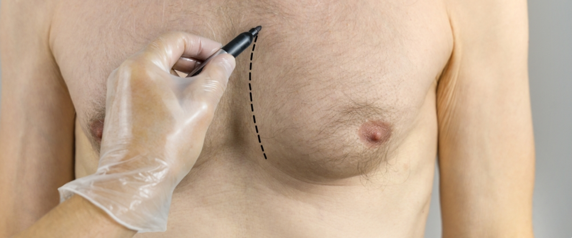
When the gland has to come out, it is cut out rather than aspirated. The standard route is an incision along the lower border of the areola — a periareolar or hemiareolar approach — placed at the junction between pigmented and unpigmented skin so the scar has somewhere to hide. The Breast Care review notes that for milder presentations, a periareolar edge incision to subcutaneous mastectomy can suffice on its own.
The trade-off is not free. A review of scar-minimizing techniques published in JPRAS Open cites a comparison of periareolar and submammary incisions in which satisfaction with chest and scar appearance did not differ significantly, but periareolar incisions carried a statistically significantly higher complication rate.
Liposuction-assisted excision
Most contemporary practice is not one technique or the other but both in sequence. The JPRAS Open review notes that many authors combine sharp glandular resection with liposuction specifically to homogenize the result: suction handles the diffuse fat and blends the edges, while sharp resection handles the disc that suction cannot. StatPearls adds that ultrasound-assisted liposuction outperforms standard suction-assisted liposuction in treating gynecomastia.
The pull-through technique
A middle option exists. The Breast Care review describes a scar-reducing approach in which liposuction is combined with a pull-through maneuver: the gland is grasped with a clamp through the existing liposuction access and drawn out, without an additional incision. The review names the constraint directly — excess skin is the limiting factor. It suits a modest disc and a skin envelope expected to retract, not a chest with significant redundancy.
Simon Grading and What Each Grade Usually Implies
Surgeons need shared language for how much chest there is and whether the skin will cooperate. Simon's classification, reproduced in the JPRAS Open review, does that in four steps.
- Grade I — small breast enlargement, no skin excess.
- Grade IIa — moderate enlargement, no skin excess.
- Grade IIb — moderate enlargement with skin excess.
- Grade III — marked enlargement with skin excess.
The break point is between IIa and IIb. The review states that from grade IIb onward the surgeon is confronted with skin excess, and that grades IIb and III are classically candidates for skin resection. StatPearls maps technique onto those grades similarly: grade I and grade IIa can be treated with liposuction and surgical excision, while grade IIb calls for open excision with possible skin resection where significant ptosis is present.
Treat this as a way of describing a chest, not an algorithm. The Breast Care review is candid that the evidence is thin and that no recommendation can be made as to a single best surgical approach.
When Skin Redundancy Enters the Plan
Skin is the second axis of the decision, independent of the fat-versus-gland question. Remove enough volume from an envelope that will not shrink and you trade a full chest for a loose one. There is active interest in avoiding skin excision altogether. The JPRAS Open review identified studies of laser-, ultrasound- and radiofrequency-assisted liposuction, microneedling, external plaster lifting of the nipple-areola complex, and transcutaneous netting as ways of encouraging skin retraction without a resection scar. Complication rates in those series ranged from 1.5 to 10 percent, but the review concludes that controlled studies are lacking and that skin resection is still often performed where redundancy is high.
When skin does have to come out, the pattern determines the scar — a periareolar purse-string resection for limited excess, a lateral or boomerang pattern, or a transverse resection with free nipple grafting for abundant excess. None is a small addition, which is why skin belongs in the plan before surgery rather than being discovered during it.
Over-Resection, Saucer Deformity and Nipple Position
The most instructive complication in this field is caused by removing too much rather than too little. Take out all of the tissue directly beneath the areola and the skin there has nothing to sit on. The result is a depression — described in the surgical literature as a saucer or crater deformity — in which the areola sinks below the level of the surrounding chest and often tethers to the muscle beneath. It is visible in raking light and in photographs, and it is considerably harder to correct than a residual bump.
Prevention is a specific technical point. The JPRAS Open review, describing its authors' own infra-areolar approach, states the rule as leaving a disc of gland roughly half a centimeter thick behind the nipple-areola complex to avoid necrosis, nipple retraction and areolar dimpling. A small, deliberate remnant is part of a correct operation, not a failure of one.
Suction produces its own version of the problem: StatPearls notes that visible skin dimpling during liposuction means the cannula is running too superficially, and lists contour deformities as the most frequent complication of liposuction overall. Nipple position belongs in the same conversation, since ASPS notes excision is required if the areola is to be reduced or the nipple moved.
What Each Approach Actually Leaves Behind
This is the comparison worth carrying into a consultation.
Liposuction alone leaves the glandular disc in place, leaves the skin envelope to retract on its own, and leaves only small access scars. It is right for a fat-dominant chest and wrong everywhere else, where it produces a flatter periphery around a bump that is now easier to see and feel.
Excision alone removes the disc but leaves surrounding fat untouched, which can produce a step where the resected area meets the fat around it. Isolated sharp resection is therefore uncommon outside small, purely glandular cases.
Liposuction with excision removes both components and lets the surgeon blend the two zones, at the cost of an areolar incision. It is the most commonly described plan for mixed presentations.
Any of the three plus skin excision adds a longer scar and, in some patterns, repositioning or grafting of the nipple-areola complex, in exchange for a tighter envelope.
Ask which of these four is being proposed, and what will still be palpable under the areola once healing is complete.
The Honest Complication Set
ASPS publishes the risks of gynecomastia surgery for patients: anesthesia risks, bleeding or hematoma, blood clots, breast asymmetry, contour and shape irregularities, changes in nipple or breast sensation that may be temporary or permanent, fluid accumulation or seroma, fat necrosis, infection, persistent pain, poor wound healing, unfavorable scarring, and possible revision surgery.
Two of those deserve numbers. The JPRAS Open review states that the most common major complication is hematoma, at an average reported rate of 5.8 percent — which is why compression, drainage and quilting techniques feature so heavily in surgical discussion. Seroma, a collection of tissue fluid rather than blood, is listed by StatPearls among the common complications of liposuction generally.
Asymmetry deserves framing rather than a number. Two sides of a chest are rarely identical beforehand, are rarely graded identically, and do not heal at identical rates. Across the series in the JPRAS Open review, satisfaction after surgical correction averaged around 84.5 percent — a good result to plan for, and not a guarantee.
When a Chest Needs a Medical Workup, Not a Cosmetic Plan
Some findings should stop a cosmetic conversation until a physician has assessed them: enlargement on one side only, enlargement that appeared or grew quickly, a chest that is tender or painful, a lump that feels hard rather than rubbery, a mass sitting off-center from the nipple, or one that seems fixed to skin or deeper tissue. So do nipple discharge, nipple retraction, skin dimpling, ulceration, scaly or reddened skin over the areola, and swollen lymph nodes in the armpit. The National Cancer Institute lists all of these among the signs of breast cancer in men.
Male breast cancer is uncommon. NCI states that fewer than one in a hundred breast cancers in the United States occur in men, and MedlinePlus notes it happens most often between the ages of 60 and 70. Uncommon is not impossible, and the Breast Care review is unambiguous that malignant change must always be ruled out, with core biopsy where there is doubt. Because routine screening mammography is not generally recommended for men, NCI describes the diagnostic pathway as clinical examination, ultrasound and mammogram, and usually a biopsy.
The Breast Care review also describes standard laboratory testing alongside examination of the testes. StatPearls, by contrast, notes that healthy men with long-standing gynecomastia of more than twelve months and no abnormal findings need no further workup. The difference between those positions is duration and physical findings.
Settling the Decision Before the Operation
A plan made in the operating room is a plan you did not consent to. Before surgery, you should be able to state the following in your own words: which of the four approaches is planned; where the incisions will be; whether skin is being excised, and in what pattern; whether the nipple-areola complex is being reduced, moved or grafted; what is deliberately being left beneath the areola; and what happens if the tissue turns out more glandular than the examination suggested. Ask to be examined sitting and lying down, because skin behaves differently in each position.
What YONSEI LEEDS CLINIC States About Its Approach
YONSEI LEEDS CLINIC is a plastic surgery and aesthetic medicine clinic in Seocho-gu, Seoul, led by Dr. In-Ho Khang, who holds an MD from Yonsei University College of Medicine and serves as Chief Academic Director of Plastic Surgery at the Korean Society of Aesthetic Surgery & Laser. The clinic identifies gynecomastia correction as one of its highest-volume specialties and lists it first among the procedures U.S. patients request.
On the specific question this article is about, the clinic's published position matches the technique logic above. It states that gynecomastia may involve firm glandular tissue beneath the nipple, excess subcutaneous fat, or both; that glandular tissue does not reliably respond to exercise or weight management; and that treatment may accordingly involve glandular excision, liposuction-based fat reduction, or a combination, depending on the patient's anatomy. It states that Dr. Khang personally evaluates each chest and distinguishes glandular, fatty and mixed presentations before selecting the approach, and that he conducts the consultation, the surgery and the post-operative assessment. It describes the goal as a chest contour that is flat, natural and symmetrical.
The clinic describes Dr. Khang as a board-certified specialist without naming the issuing board, so the practical step is to ask to see the certificate at consultation. His background appears on the clinic's doctor profile page, and inquiries and online consultations run through its contact page.
The post-surgery care program the clinic publishes
The clinic publishes a staged chest recovery protocol rather than a single discharge sheet: assessment of the wound, garment fit and swelling in the first days; lymphatic and swelling support with compression and activity guidance over the first two weeks; scar management from around weeks two to four once the incision has closed adequately; radiofrequency-based skin firmness care from roughly weeks four to eight where clinically appropriate; a mid-recovery evaluation around weeks six to eight; and a final review of scar maturation, skin firmness and contour symmetry at months three to six. It states that visible swelling commonly decreases substantially over about four to eight weeks and that scars may continue improving over three to six months, while adding that healing varies by individual. It also states in its own documentation that it should not claim guaranteed results, guaranteed symmetry, specific success rates, complication-free outcomes or exact recovery timelines.
What is not published
Naming the gaps is more useful than filling them. The clinic does not publish an anesthesia protocol for gynecomastia surgery, a procedure-specific complication list, a standard operative time, case volumes, years in practice, booking lead times, revision policy specifics, or payment arrangements. Its published equipment descriptions — including the EVA-assisted system with AIRS technology and ADDIT technology — sit under its liposuction-by-area service, and it does not publish which of these it uses in gynecomastia cases. Ask directly.
Planning It From the United States
Seoul runs roughly 13 to 17 hours ahead of the continental United States, so the pre-arrival teleconsultation the clinic states it offers — along with written pre-operative documentation and remote follow-up after you return home — usually means an early or late call.
Two travel points are specific to chest surgery. The CDC's Yellow Book advises medical tourists not to fly for ten days after chest or abdominal surgery because of the pressure changes involved, and notes that air travel and surgery each raise the risk of blood clots. The same guidance recommends arranging follow-up care before you travel, requesting copies of your records in English, and being aware that devices and medicines used abroad are not necessarily subject to the same regulatory oversight as in the United States. The clinic states it provides individual guidance on length of stay and on the timing of the initial post-operative assessment; ask for that in writing before booking flights.
The Bottom Line
- Liposuction removes fat and does not reliably remove fibrous glandular tissue, so a glandular or mixed chest treated by suction alone will usually retain a palpable disc beneath the areola.
- Suction alone is the correct, complete operation for a genuinely fat-dominant chest with no subareolar mass — and the wrong one everywhere else.
- Simon grading describes volume and skin excess in four steps; the break point is grade IIb, from which skin resection classically enters the plan, though published guidance stops short of naming one best technique.
- Leaving a deliberate thin disc of gland behind the nipple-areola complex is part of a correct excision; removing all of it risks a saucer deformity that is harder to correct than a residual bump.
- Hematoma is the most commonly reported major complication, at an average of about 5.8 percent, and unilateral, rapidly progressive or tender enlargement should be medically evaluated before any cosmetic plan.
Frequently Asked Questions
Can liposuction alone get rid of gynecomastia?
Only if the enlargement is fat rather than gland. The American Society of Plastic Surgeons states that liposuction alone may be used where gynecomastia is primarily the result of excess fatty tissue, and that excision is recommended where glandular tissue must be removed. StatPearls notes that in chronic cases the tissue becomes fibrotic and liposuction may not work, so an open procedure may be required.
Will an examination tell the difference, or do I need a scan?
Examination usually does most of the work. Published guidance states that palpation should distinguish fatty tissue from glandular tissue clinically, alongside assessment of skin excess, ptosis and nipple retraction. Ultrasound and, where indicated, mammography confirm the finding or investigate anything that does not behave like ordinary gynecomastia.
Why would I still need an areolar incision if I am also having liposuction?
Because the two instruments do different jobs. Suction blends surrounding fat and reduces volume, but the fibrous disc under the nipple is generally removed sharply, and that requires access. An incision at the border of the areola places the scar at the junction between pigmented and unpigmented skin. Some surgeons instead draw the gland out through the liposuction access, though skin excess limits when that is suitable.
What is a saucer or crater deformity?
It is a depression under the areola caused by removing too much tissue beneath the nipple-areola complex, leaving the skin there with nothing to sit on. Surgical literature describes leaving a disc of gland roughly half a centimeter thick behind the complex to avoid necrosis, nipple retraction and areolar dimpling. Correcting an over-resection is generally harder than removing a small remnant later.
What is the most common complication of gynecomastia surgery?
A published review reports hematoma — a collection of blood in the surgical space — as the most common major complication, at an average rate of about 5.8 percent. ASPS also lists seroma, asymmetry, contour irregularities, changes in nipple sensation, infection, unfavorable scarring and possible revision surgery among the risks patients should be counseled about.
When should I see a doctor rather than book cosmetic surgery?
If the enlargement is on one side only, has grown quickly, is painful, feels hard, sits off-center from the nipple, or comes with nipple discharge, nipple retraction, skin dimpling, ulceration or swollen lymph nodes in the armpit. The National Cancer Institute lists these among the signs of breast cancer in men, and surgical guidance treats ruling out malignancy as a standing requirement.
This article is for general informational purposes and does not constitute individualized medical advice, and it is not a self-diagnosis tool. Surgical candidacy, technique, and expected outcomes can only be determined through a consultation with a qualified plastic surgeon.
More Articles
Continue reading our clinical guides.

Why Korea Body Contouring Prices Stay Low Despite Global Inflation Rise
While global inflation has driven up the cost of surgical procedures in many Western markets, Korean body contouring clinics have maintained stable pricing through efficiency and modern liposuction protocols.
Learn More

Breast Fat Grafting Costs: Korea vs Japan Price Comparison 2026
Autologous breast fat grafting uses a patient's own fat cells to enhance breast volume and shape. Compare 2026 breast fat grafting costs and SVF systems in South Korea vs Japan.
Learn More

Body Lift Efficacy: 90% Satisfaction in Korean Reconstruction Studies
The human body undergoes profound structural modifications following significant weight loss, childbirth, or natural structural aging. Learn how high-precision body lift surgery in Korea achieves 90% patient satisfaction.
Learn More